
![]()
Reader, I have a bit of a visual treat for you today. A group at the Heidelberg branch of the European Molecular Biology Lab (EMBL) published a paper in Science a few months ago, detailing a full microscopic scan of the first 24 hours in the development of the zebrafish embryo, from a handful of cells to when the first structuring starts to occur. And, in doing so, they produced some startlingly beautiful videos of the first moments of life.
Imaging an Embryo
Their method is a scientific tour-de-force. They produce a high resolution 3-D image (1500x1500x350 pixels) every 90 seconds; and they do this without harming the fish at all, so it stays alive and perfectly happy, developing normally. The way they do this is by something that they call ŌĆ£Digital scanned laser light sheet fluorescence microscopyŌĆØ (DSLM), which in essence involves modifying the embryo to make a protein in their nuclei that glows green under lazer light (the GFP protein). They then scan the embryo with a sheet of lazer light, and detect how the protein glows. They introduce lots of fancy new tricks, which you can read about here, but I wonŌĆÖt go into detail (perhaps because I donŌĆÖt really understand it).
As IŌĆÖve already mentioned, the most striking thing about this paper is the videos they produce. The video below shows the embryo developing from two different angles (click once to play, and click again for a higher resolution video):
(this looks a little like a fish-bowl full of firefliesŌĆ”)
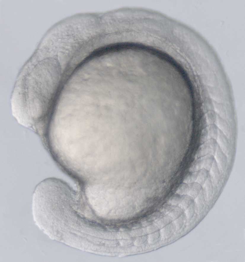
The amount of detail you can see here is startling. The white blobs are individual nuclei, each in itŌĆÖs own cell. You can see them dividing, in little bursts of activity at first, but soon it gets chaotic. Then you can see the cells migrate around the embryo, making a ball of cells, and then when this is done they start forming the ring structure, with the head bulge, thatŌĆÖll late become the fetus (this is what it looks like a bit later on).
Digitizing an Embryo
Just getting images with this resolution and detail is remarkable. However, they werenŌĆÖt finished. They used a set of algorithms to scan through every image, and digitally record the location of every nuclei, and track the nucleiŌĆÖs movement over time. They went from having 3D images of the embryo, to having a digital 3D model that they could manipulate at will. For instance, they can figure out how fast each cell is moving at any given time:
Orange nuclei are moving faster; you can see that different portions are active at different times; some regions are full of shifting nuclei, while others are still. However, they is more complex analysis you can do. The authors used their digital embryo to look at how the embryo goes from being an undifferentiated ball to being an asymmetric fetus, and to see how different types of tissue form (these have been looked into before, and the answers were already known in general terms, but the digital embryo allowed detailed examination).
One nice thing they can do is that they digitally mark cells in the portion of the embryo that will become a particular organ (in this case the eye, which is easy to find at 24 hours), and trace these nuclei back through time to see where they came from. This video shows the nuclei that become the eye in red:
I will stop with the videos now, but you can download all 16 lovingly rendered films, showing a range of different lines of investigation, from here.
A Digital Resource
The digital embryo is an impressive technological achievement, and being able to visualise development in such a striking and beautiful way is an end in itself. However, the Science paper doesnŌĆÖt really tell us anything we didnŌĆÖt already know about vertebrate development; it investigates a few questions, and gives some interesting tidbits, but these feel underwhelming compared to how impressive the technology is.
This is less disheartening than it might seem, and is typical of this kind of work. To take an extreme example, the human genome papers give some interesting observations about human genetics, but these were dwarfed by the scale of the project itself. Ultimately, this paper is a promise of science to come. The datasets (a terrifying 3 terabytes per embryo) can be used by other scientists for more detailed investigations into systems biology, hopefully being used to test out models of early development. The technology is ready to put in pretty much any embryo, of virtually any species, including developmental mutants. When we have screened a load of different species and mutations, we can start asking exactly WHEN do the differences between them arise, WHERE and hopefully HOW. And, while DLSM currently looks only at where nuclei are, there is no reason that this technology couldnŌĆÖt be modified to look for cells that express a particular protein, and even measure the amount of that protein in each cell; then we can start looking at what roles different genes are playing, and how they interact with each other.
Developmental biology is becoming increasingly digital; a field that consisted of squinting down a microscope all day is now full of complex quantitative data (like the FlyEx database for Drosophila), and new models and mathematical methods have had to be developed to make use of these resources. And no one person can develop these tools; it is only when experimental and theoretical biologists, along with engineers, computer scientists and mathematicians, come together that people can make any sense of it all.
I got most of my information about this from Gareth Powell at the Cell Surface Signaling Laboratory at the Sanger Institute
Keller, P., Schmidt, A., Wittbrodt, J., & Stelzer, E. (2008). Reconstruction of Zebrafish Early Embryonic Development by Scanned Light Sheet Microscopy Science, 322 (5904), 1065-1069 DOI: 10.1126/science.1162493


Why is there an ŌĆśanimalŌĆÖ and a ŌĆśvegetalŌĆÖ pole, instead of more isotropic division rates?
I think it is mostly a mechanism of producing asymmetry (which is put there by the mother); in mammals, the animal pole becomes the fetus, and the vegetal pole becomes the placenta, and you need a way of determining which will be which. Unless you want a spherical baby/placenta hybrid. Do you, do you Olaf? Do you want a spherical baby/placenta hybrid? I thought not.
Whether cells are in the vegetal or animal pole also determines what cell types they will become in the fetus, with cells in the vegetal pole mostly becoming the digestive system, and the cells in the animal pole becoming skin and bones and so on.
Wow, that is amazing. As far as not producing too many ŌĆśnewŌĆÖ discoveries goes, it is probable (especially if this new stuff has just been made) that at the moment they are testing their method, rather than trying to find new information. It does seem to have a huge potential though; do you know if itŌĆÖs been tried in any other model organisms?
Not yet, but I think they are planning to try it on chicken in the near future.